Torn Retinaculum Ankle - Peroneal Tendonitis Footeducation : A retinaculum (plural retinacula) is a band of thickened deep fascia around tendons that holds them in place.
Torn Retinaculum Ankle - Peroneal Tendonitis Footeducation : A retinaculum (plural retinacula) is a band of thickened deep fascia around tendons that holds them in place.. Attention was then directed to the medial aspecto fo the right ankle joint, where. Again a previously torn retinaculum will be thickened and ill defined compared to a normal retinaculum which is usually thin and well defined. Flexor retinaculum at the ankle is formed by reinforcement of the deep fascia of the leg by transverse collagen bundles and functions to prevent 'bowstringing' of tendons as they pass the tibiotalar joint. Ankle ext retinaculum torn after fasciotomy & injury in pt. Patient had an ankle injury and the retinaculum over the peroneal brevis and longus was torn.
Inferior extensor retinaculum pain or strain can also be accompanied with swelling and tenderness in the ankle region of the foot and the lower legs. Previously torn extensor retinaculum of ankle which is now markedly thickened and irregular (blue arrows). This is called an avulsion fracture. Your doctor may also order a magnetic resonance imaging (mri) scan of your ankle. Retinaculum also acts as a pulley system increasing mechanical advantage.
Ankle Stability Retinaculum Connection from static.wixstatic.com A retinaculum (plural retinacula) is a band of thickened deep fascia around tendons that holds them in place. Retinaculum repair is gaining popularity. 43 years experience general surgery. This is called an avulsion fracture. In surgery to repair the retinaculum, the surgeon first makes an incision along the back and lower edge of the fibula bone. The main symptoms of inferior extensor retinaculum pain are: Here's the meat of the op report: Any damaged tendon is removed prior to repair.
Depending on the severity of tear, sometimes surgery is needed to repair the retinaculum.
Your doctor may also order a magnetic resonance imaging (mri) scan of your ankle. My podiatrist fixed a torn flexor retinaculum of the rt ankle. Forced ankle eversion and forced dorsiflexion with a contracted tibialis posterior tendon is the most common mechanism of tendon subluxation, though the phenomenon is also seen with medial malleolar fractures.medial flexor retinaculum injuries are also usually associated with deltoid ligament injuries. I need another person's input. T he retinacula of the ankle are distinct structures defined as regions of localized thickening of the superficial aponeurosis covering the deep structures of the distal portion of the leg, ankle, and foot. Tibialis anterior tendon (arrowheads) is most medially located and passes though tunnels formed by oblique superomedial limb (short arrows) and oblique inferomedial limb (long arrows) of. In this article, we present the assessment, diagnostic algorithm and a new therapeutic option for the distal dislocation of the long peroneal tendon due to isolated inferior peroneal retinaculum (ipr) tear. Here's the meat of the op report: The retinacula of the ankle are regions of localized thickening of superficial aponeurosis that provide mechanical strength to prevent tendon bowstringing. This procedure restores the normal anatomy of the retinaculum that covers and reinforces the tendon sheath around the peroneal tendons. He came back after 2 weeks and reported that he had a left ankle injury when playing basketball and was using crutches and was on air boots when came to see me. Previously torn extensor retinaculum of ankle which is now markedly thickened and irregular (blue arrows). The gel is used by rubbing it at the injured site up to the ankle.
Between 2001 and 2011 three patients with distal peroneal tendon dislocation were operated. He said that the temporary insoles were ok to ease the pain in the bottom of the foot but achille's tendon still hurting and pain on the lateral border hasn't changed. Patient had an ankle injury and the retinaculum over the peroneal brevis and longus was torn. Once the cast is off, a strengthening program is prescribed by a physiotherapist to help you return back to normal function. Skin is closed with suture, followed by application of bandages and below the knee cast
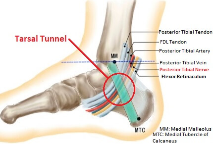
Tarsal Tunnel Syndrome Causes Symptoms Treatment from www.foot-pain-explored.com It is not part of any muscle. Tearing of retinacula is more commonly seen at the ankle. Once the cast is off, a strengthening program is prescribed by a physiotherapist to help you return back to normal function. I'm looking at coding it with cpt code 28200. This is called an avulsion fracture. The retinaculum ligament is then repaired and advanced back to the point of original attachment. The physician repaired the retinaculum which holds these down. The tear is repaired with suture above.
Your doctor may also order a magnetic resonance imaging (mri) scan of your ankle.
Occasionally, the covering that holds the peroneus tendons behind the lateral malleolus (the retinaculum) can be loose or torn and the tendons can snap back and forth out of their normal grooves, this snapping sensation is felt by the patient and can causes further stress/friction on the tendons. Retinaculum injuries are the most common, followed by type 3. I'm looking at coding it with cpt code 28200. The retinaculum ligament is then repaired and advanced back to the point of original attachment. Your doctor may also order a magnetic resonance imaging (mri) scan of your ankle. Ankle ext retinaculum torn after fasciotomy & injury in pt. He came back after 2 weeks and reported that he had a left ankle injury when playing basketball and was using crutches and was on air boots when came to see me. The symptoms of subluxation may include: It is not part of any muscle. However, the physician wants to bill for repair of dislocating tendon (27675) but in addition wants to bill for repairing the tendons themselves (27659) which i don't understand. This is called an avulsion fracture. Retinaculum repair is a procedure designed to restore the retinaculum ligament, the bands of tissue that surround and stabilize the peroneal tendons. T he retinacula of the ankle are distinct structures defined as regions of localized thickening of the superficial aponeurosis covering the deep structures of the distal portion of the leg, ankle, and foot.
Previously torn extensor retinaculum of ankle which is now markedly thickened and irregular (blue arrows). Here's the meat of the op report: The retinaculum ligament is then repaired and advanced back to the point of original attachment. I'm looking at coding it with cpt code 28200. Due to the mechanism of injury, superior peroneal retinaculum tears usually go misdiagnosed as just a lateral ankle sprain.
Snapping Ankle Or Clicking Ankle Core Concepts Physiotherapy from www.coreconcepts.com.sg Inferior extensor retinaculum pain or strain can also be accompanied with swelling and tenderness in the ankle region of the foot and the lower legs. During this procedure, an incision is made near the back and outer edge of the fibula (ankle bone). I need another person's input. Forced ankle eversion and forced dorsiflexion with a contracted tibialis posterior tendon is the most common mechanism of tendon subluxation, though the phenomenon is also seen with medial malleolar fractures.medial flexor retinaculum injuries are also usually associated with deltoid ligament injuries. After surgery, a cast or boot is used to help stabilise the ankle and allow the new tissue to heal successfully. Retinaculum repair is gaining popularity. Attention was then directed to the medial aspecto fo the right ankle joint, where. Retinaculum injuries are the most common, followed by type 3.
The tear is repaired with suture above.
He came back after 2 weeks and reported that he had a left ankle injury when playing basketball and was using crutches and was on air boots when came to see me. During this procedure, an incision is made near the back and outer edge of the fibula (ankle bone). Previously torn extensor retinaculum of ankle which is now markedly thickened and irregular (blue arrows). Acute, limited tears of a single peroneal tendon may be debrided and repaired. The retinacula of the ankle are regions of localized thickening of superficial aponeurosis that provide mechanical strength to prevent tendon bowstringing. The gel is used by rubbing it at the injured site up to the ankle. Here's the meat of the op report: The podiatrist is looking at 27695. Ankle ext retinaculum torn after fasciotomy & injury in pt. Additionally, what does the retinaculum do? The main symptoms of inferior extensor retinaculum pain are: Plantaris tendon (if you have one) overlay may do the. A retinaculum (plural retinacula) is a band of thickened deep fascia around tendons that holds them in place.


Posting Komentar
0 Komentar